Home

inflasi Pygmalion Dislokasi green marker mitochondria facs berjaya Menggunakan Apa yang salah

Cellular Allometry of Mitochondrial Functionality Establishes the Optimal  Cell Size - ScienceDirect
Cellular Allometry of Mitochondrial Functionality Establishes the Optimal Cell Size - ScienceDirect

Comparison of JC‐1 and MitoTracker probes for mitochondrial viability  assessment in stored canine platelet concentrates: A flow cytometry study -  Marcondes - 2019 - Cytometry Part A - Wiley Online Library
Comparison of JC‐1 and MitoTracker probes for mitochondrial viability assessment in stored canine platelet concentrates: A flow cytometry study - Marcondes - 2019 - Cytometry Part A - Wiley Online Library

Loss of Mitochondrial Function Impairs Lysosomes* - Journal of Biological  Chemistry
Loss of Mitochondrial Function Impairs Lysosomes* - Journal of Biological Chemistry

Mitotracker Green Fm (Thermo Fisher) | Bioz | Ratings For Life-Science  Research
Mitotracker Green Fm (Thermo Fisher) | Bioz | Ratings For Life-Science Research

Mitochondrial dysfunction in Top1mt2/2 cells. FACS analysis of ROS (A),...  | Download Scientific Diagram
Mitochondrial dysfunction in Top1mt2/2 cells. FACS analysis of ROS (A),... | Download Scientific Diagram

The plot of JC-1 red verse green emission from flow cytometry on... |  Download Scientific Diagram
The plot of JC-1 red verse green emission from flow cytometry on... | Download Scientific Diagram

Cytometric assessment of mitochondria using fluorescent probes -  Cottet‐Rousselle - 2011 - Cytometry Part A - Wiley Online Library
Cytometric assessment of mitochondria using fluorescent probes - Cottet‐Rousselle - 2011 - Cytometry Part A - Wiley Online Library

Dysfunctional mitochondria accumulate in Atg5-deficient MEFs. (A and B)...  | Download Scientific Diagram
Dysfunctional mitochondria accumulate in Atg5-deficient MEFs. (A and B)... | Download Scientific Diagram

Flow Cytometry using MitoTracker Mitochondrial dye?

Using flow cytometry for mitochondrial assays - ScienceDirect

Multicolor Flow Cytometry-based Quantification of Mitochondria and  Lysosomes in T Cells | Protocol
Multicolor Flow Cytometry-based Quantification of Mitochondria and Lysosomes in T Cells | Protocol

BioTracker 488 Green Mitochondria Dye Live cell imaging mitochondrial dye  that stains the membrane of mitochondria used to detect cell viability,  metabolic activity and overall cell health.
BioTracker 488 Green Mitochondria Dye Live cell imaging mitochondrial dye that stains the membrane of mitochondria used to detect cell viability, metabolic activity and overall cell health.

Improving the Accuracy of Flow Cytometric Assessment of Mitochondrial  Membrane Potential in Hematopoietic Stem and Progenitor Cells Through the  Inhibition of Efflux Pumps | Protocol
Improving the Accuracy of Flow Cytometric Assessment of Mitochondrial Membrane Potential in Hematopoietic Stem and Progenitor Cells Through the Inhibition of Efflux Pumps | Protocol

Flow cytometry analysis of isolated mitochondria. (A) cells were... |  Download Scientific Diagram
Flow cytometry analysis of isolated mitochondria. (A) cells were... | Download Scientific Diagram

PLOS ONE: Reduced Basal Autophagy and Impaired Mitochondrial Dynamics Due  to Loss of Parkinson's Disease-Associated Protein DJ-1
PLOS ONE: Reduced Basal Autophagy and Impaired Mitochondrial Dynamics Due to Loss of Parkinson's Disease-Associated Protein DJ-1
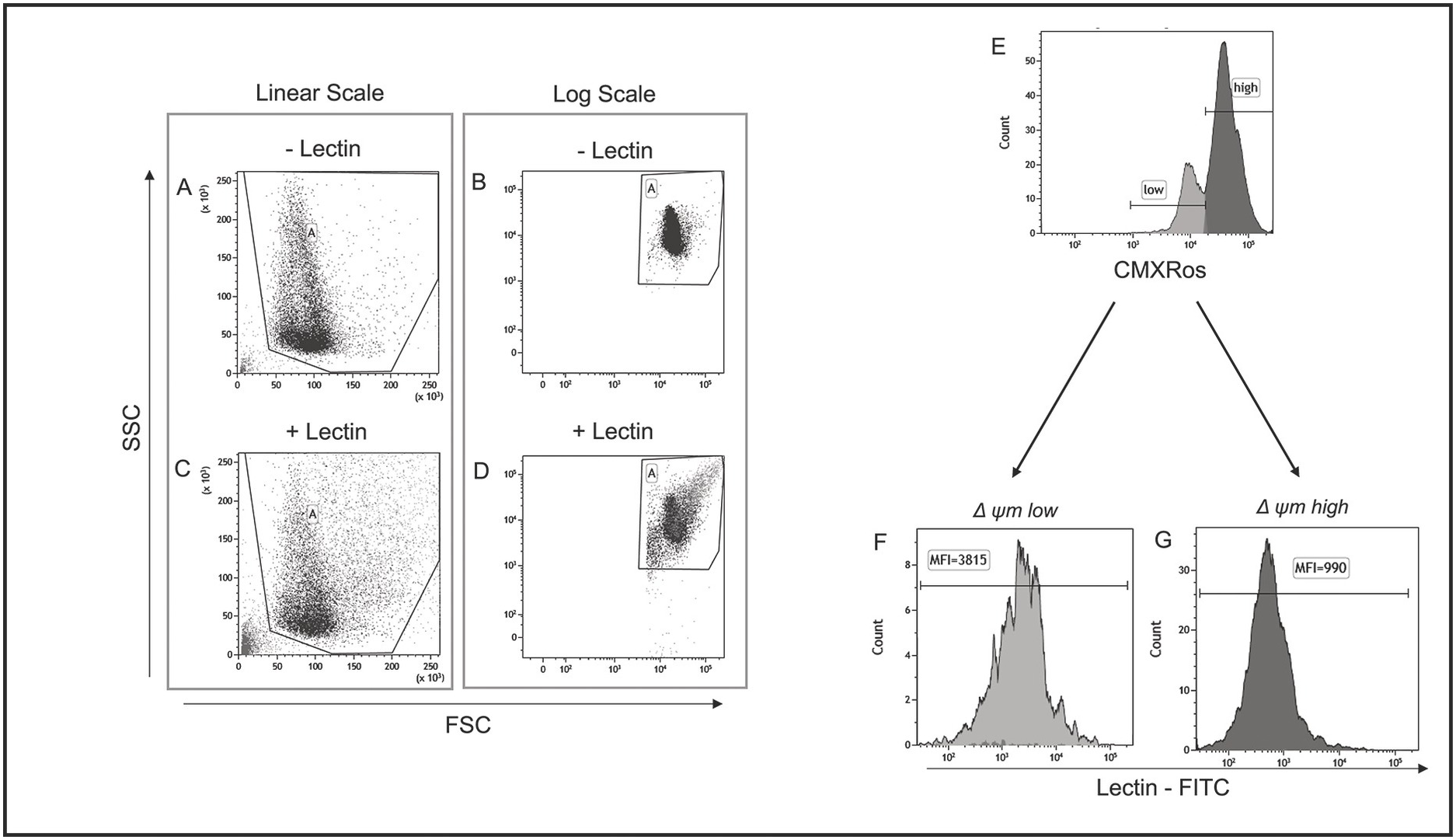
Determination of Mitochondrial Membrane Potential by Flow Cytometry in  Human Sperm Cells (Chapter 8) - Manual of Sperm Function Testing in Human  Assisted Reproduction
Determination of Mitochondrial Membrane Potential by Flow Cytometry in Human Sperm Cells (Chapter 8) - Manual of Sperm Function Testing in Human Assisted Reproduction
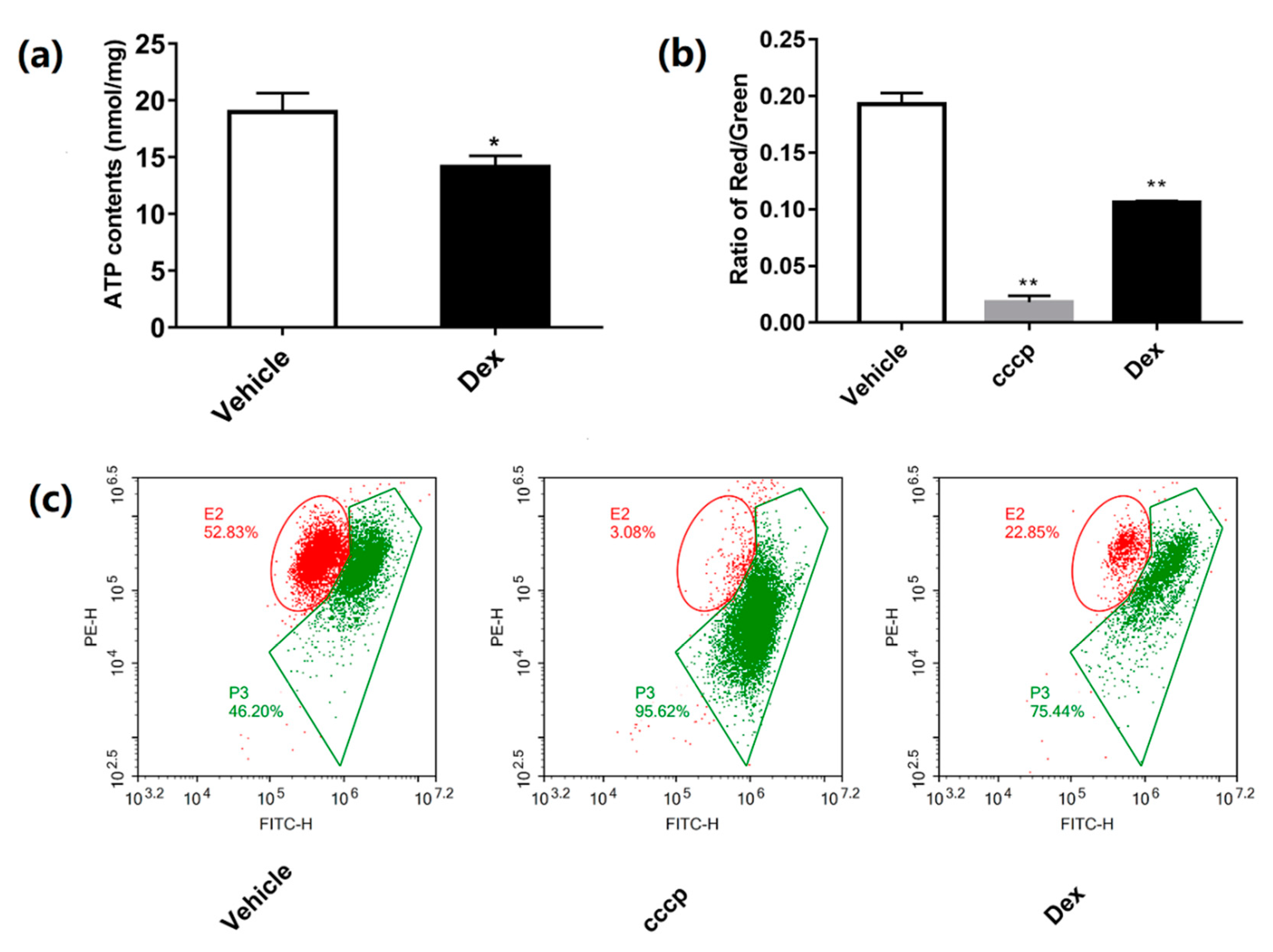
Molecules | Free Full-Text | Dexamethasone-Induced Mitochondrial  Dysfunction and Insulin Resistance-Study in 3T3-L1 Adipocytes and  Mitochondria Isolated from Mouse Liver | HTML
Molecules | Free Full-Text | Dexamethasone-Induced Mitochondrial Dysfunction and Insulin Resistance-Study in 3T3-L1 Adipocytes and Mitochondria Isolated from Mouse Liver | HTML
NIR Mitochondrial Membrane Potential Assay Kit (Flow Cytometry) (ab112149)

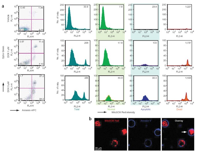
Simultaneous detection of apoptosis and mitochondrial superoxide production  in live cells by flow cytometry and confocal microscopy | Nature Protocols
Simultaneous detection of apoptosis and mitochondrial superoxide production in live cells by flow cytometry and confocal microscopy | Nature Protocols
Autophagy - Flow Cytometry Core Facility

Multicolor Flow Cytometry-based Quantification of Mitochondria and  Lysosomes in T Cells | Protocol
Multicolor Flow Cytometry-based Quantification of Mitochondria and Lysosomes in T Cells | Protocol

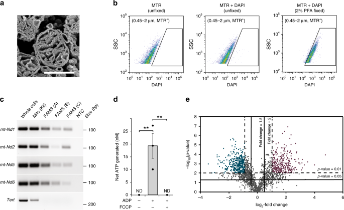
A nanoscale, multi-parametric flow cytometry-based platform to study  mitochondrial heterogeneity and mitochondrial DNA dynamics | Communications  Biology
A nanoscale, multi-parametric flow cytometry-based platform to study mitochondrial heterogeneity and mitochondrial DNA dynamics | Communications Biology

CCN6 regulates mitochondrial function

JC-10 Mitochondrial Membrane Potential Assay Kit (Flow Cytometry) (ab112133)

SCAN1-TDP1 trapping on mitochondrial DNA promotes mitochondrial dysfunction  and mitophagy | Science Advances
SCAN1-TDP1 trapping on mitochondrial DNA promotes mitochondrial dysfunction and mitophagy | Science Advances

Assessment of Mitochondrial DNA Content and Mass in ...